

Triple Arthrodesis
Indications
Triple arthrodesis is indicated for patients who have a deformity of the hindfoot, such as acquired adult flatfoot deformity, where there is arthritis or stiffness in the involved joints. This type of procedure sometimes offers a more reliable result than other hindfoot corrective procedures. Because of the importance of preserving all the joints in the foot, this procedure is done a lot less often than it used to be. However, if there is enough arthritis or dysfunction in the involved joints where preservation is improbable, then a triple arthrodesis may be effective.
Procedure
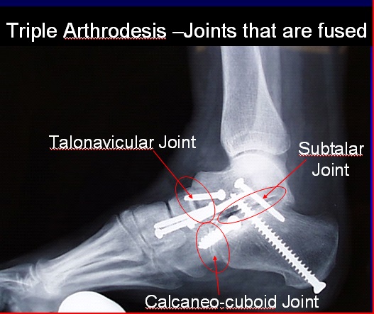
This surgical procedure involves the fusion of three joints: the talonavicular joint, the subtalar joint, and the calcaneal-cuboid joint (See Figure 1). Two incisions are made on both sides of the foot; one is made on the inside (medial) to expose the talonavicular joint, and the other is made on the outside to expose the subtalar joint, the calcaneal-cuboid joint, and the outside (lateral) aspect of the talonavicular joint. Once exposed, all of the remaining cartilage from the joints is removed and prepared for fusion. Typically, the joint is then packed with bone graft from other bones, such as the proximal tibia or the iliac crest (located at the top of the pelvic bone). Once all the joints have been prepared and the bone graft has been placed, the hindfoot position is corrected to a neutral position. Once repositioned, the foot is first provisionally fixed with wires, and then definitively fixed with screws or plates in each joint. Once everything is in place, the incisions are closed up with sutures.
Figure 1: Triple Arthrodesis

Recovery
0-6 (or 8) weeks Post-Surgery
Patients will not bear any weight on the operated leg for the first 6-8 weeks after surgery, in order for the joints to heal. However, the talonavicular joint is challenging to become fully healed, so the healing process could be longer. During this time, patients are usually in a cast or splint to immobilize the foot in its corrected position.
6 (or 8) weeks – 12 (or 14) weeks Post-Surgery
At 6 or 8 weeks postoperatively, if x-rays demonstrate satisfactory healing, patients are then allowed to begin weight-bearing as tolerated in a protective cast boot. This boot can be removed to allow showering and physical therapy. At this point physical therapy is started. Patients will work on strengthening their muscles, improving their gait, and working on their range of motion.
12 (or 14) week + Post-Surgery
At that point, patients can begin walking in a stiff soled shoe, often with a supportive ankle device.
Patients will obtain approximately 60-70% of their recovery in 5-6 months. However, it is often 15 or even 18 months before a patient reaches their point of maximal improvement.
Potential Complications
General Complications
Specific Complications
- Nonunion. A nonunion occurs when the fusion site fails to adequately knit together. The nonunion rate is dependent upon: the surgical technique; the patient’s underlying condition; whether the patient smokes cigarettes; and the patient’s compliance with the postoperative non weight-bearing protocol. The typical nonunion rate is on the order of 5% for the subtalar and the calcaneal-cuboid joint, but the nonunion rate for the talonavicular joint is around 5-8%. If a patient does have a nonunion or a delayed union, they may require a longer period of non weight-bearing and, in some instances, will require revision surgery.
- Nerve Injury. Injury to the nerve on the outside aspect of the foot (superficial peroneal nerve) can occur due to the placement of the incisions. Nerve injury can occur due to retraction, direct injury, or from scarring during the recovery process. If these nerves are injured or cut, the patient could end up with numbness or pain along the path of the nerve.
- Painful Hardware. Pain may be associated with the screws that are used to secure the joints. About 10-20% of people will need to undergo removal of the screws, usually in the subtalar joint, due to discomfort, once the bones have healed.
- Arthritis of Ankle Joint. Prior to surgery, patients have movement that occurs below the ankle joint; however after surgery, the movement below the ankle joint dissipates and goes through the ankle joint itself, meaning more force is applied in that area. After 10-15 years, patients will see arthritic changes on x-ray, but patients will not necessarily be symptomatic, although it is still a possibility.
Edited July 11, 2017
mf/4.2.18