

Flexible Flatfeet
(Pediatric Flatfoot)
Edited by Gwyneth DeVries, MD FRCSC
Summary
Some children will develop a pronounced flatfoot deformity (Figure 1). Usually this deformity does not create symptoms, although some children may complain of an ache in their arch or their calf muscles with increased activity. In most instances, the flatfoot deformity is flexible and is due to a tight outer calf muscle. Treatment of flexible feet is almost always non-surgical and includes calf stretching, activity modification, and comfortable shoes often consisting of a slight heel.
Figure 1A: Flexible Flatfoot – Front View
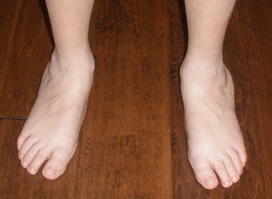

Figure 1B: Flexible Flatfoot – Side View
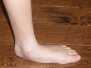

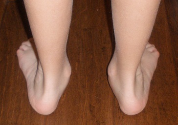
Figure 1C: Flexible Flatfoot – Back View

Is it normal for children to have flat feet?
Having flat feet is common, representing 20-30 percent of the population. Flatfoot deformities often run in families, although flat feet can also be part of normal development in any child. In most cases, as a child grows, the feet become less flat-looking as muscles become stronger. Most children with flat feet have no symptoms. In most cases, the family doctor or the other primary care provider can identify which type of flat foot is no cause for concern, and recommend a “wait and see” approach.
On the other hand, some children have quite marked flatfeet (often as adolescents) that are characterized by a striking flattening of the arch and a splaying outwards of the feet. Most of these individuals have no symptoms or may have some general aching in their arches if they are particularly active. The extent of their deformity often causes parents to be quite alarmed and seek a more specialized opinion. A careful examination will help to answer questions and guide investigations or treatment
Flexible Flatfeet versus Rigid Flatfeet
It is important from a treatment perspective to determine the type of flatfoot – either flexible or rigid. Flexible flatfeet are much more common than rigid flatfeet. Flexible flatfeet may have very flexible joints throughout the foot and are often associated with a flatfeet. Flexible flatfeet may have very flexible joints throughout the foot and are often associated with a tight calf muscle. Rigid flatfeet are usually caused by a tarsal coalition (two or more bones joined together during development, usually in the “hindfoot” bones just below the ankle joint).
Physical Examination
Children with flatfeet will have noticeable flattening of the arch on the inside of the foot. Often the foot will also be splayed outwardly (abducted). Walking will often be normal, though a limp may be noted if there is pain. Sensation to the foot will typically be normal. The physician will then try to determine whether the patient’s flatfeet are flexible (good hindfoot motion) or rigid (limited hindfoot motion). The motion of the hindfoot – specifically whether the foot can be moved inward (inversion) or outward (eversion) will determine whether the flatfoot deformity is flexible or rigid. With the child seated, the examiner will support the ankle and then gently try to move the heel from side to side. For flexible flat feet, the heel will move easily in both directions, in a rigid foot, the heel will be difficult to move in one or both directions. A “jack test” can also be used to check for flexibility. During a “jack test,” the examiner holds the heel steady and bends the big toe upwards. If an arch forms, the flatfoot is deemed ‘flexible.’ If the foot remains flat, it is rigid.
Patients with a flexible flatfoot deformity often have a very tight outer calf muscle (gastrocnemius). The examiner will test the tightness of the calf muscle by observing the range of motion of the ankle with the knee bent and with the knee straight. When the knee is bent, the calf muscle is a bit more relaxed, so the foot can be moved quite easily toward the shin bone. The angle of the foot and the lower leg is recorded as “dorsiflexion past 90 degrees” if the foot can lift past a right angle. When the same test is repeated, this time with the knee straight, then the calf muscle (gastrocnemius) is tighter, and as the foot is brought up with the knee straight, the foot will often be 20-30 degrees short of neutral (right angle to the lower leg). When this is seen in the examination setting, it is known that the calf muscle tightness occurs during normal walking (or other activities). The tight muscle prevents the ankle from bending far enough toward the shin, so extra movement takes place in the joint of the foot instead. (The part of the foot that must adapt is the “transverse tarsal region, made up of the calcaneocuboid and talonavicular joints). Extra movement through this area does allow for extra upward motion of the foot, but the forces are such that the foot also splays out to the side, creating the flatfoot deformity and sometimes leading to a stretching or painful sensation along the inside of the ankle. Having a flexible flatfoot deformity tends to concentrate increased load through the inside of the arch, the ankle, and the Achilles tendon, so it is not uncommon for children to have some symptoms in these areas, especially if they have periods of increased activity.
Imaging Studies
In both types of flatfoot (flexible or rigid) plain weight bearing x-rays of the foot will demonstrate a flattened arch. In patients with a tarsal coalition, the fused bones can often be seen on plain x-rays although it may be necessary to order a CT scan to fully assess whether a tarsal coalition is present. In some patients with vague symptoms, an MRI may help identify areas of bone edema (increased blood flow) or other potential sources of pain.
Treatment
Flexible flatfeet are almost always treated non-operatively. Treatment is not designed to change the shape of the foot, but rather to manage painful symptoms or improve overall functions for the longterm. Treatments may include:
- Activity modification: Sometimes a short (1-2 week) period of limited activities can be enough to allow any painful symptoms to settle. Less repetitive loading through tendons and joints will have the effect of decreasing irritation to these areas. In addition, a switch to activities that require less loading (ex. from running to cycling) may also help symptoms to settle.
- Calf stretching: Calf stretching is an important component of treating flexible flatfeet. It can be very helpful to try and stretch out the tight outer calf muscle, by turning the foot inward and stretching the calf with the knee straight. Working with a physical therapist can help to determine the best stretches for an individual. Stretches should be performed on a daily basis for the best long-term effect.
- Comfort shoes with a slight heel: Wearing shoes with a slight heel can be helpful. Comfort shoes can help to disperse the force up the leg more smoothly, while a slight heel can have the effect of decreasing the amount of flattening of the arch during normal standing, walking, or activity.
- Shoes and Shoe inserts: It is often guessed that individuals with a flatfoot would benefit from an orthotic that helps to “prop up” their arch. In fact, a stiff orthotic designed to elevate the arch may be uncomfortable, as the affected bones cannot actually be “moved.” However, a soft (accommodative) orthotic that can support the arch can be helpful. These orthotic inserts can often be obtained at sports stores or outdoor stores. Although a custom molded version, again paying attention to moderate, cushioning effect can be the preference of those who can manage the added expense. Replacement of any insoles or orthotics should keep pace with the shoe size and overall growth. Shoe type is up to the individual, care should be taken to decrease pressure areas and if possible, include durable materials that support the sole and the sides of the feet. This allows for selection of a variety of shoes for all weather types; even certain sandals will have these features. High top or ankle-supporting shoes are optional, not necessary, but may the preference of some individuals in certain activities.
Operative Treatment
Flexible flatfeet rarely require surgical intervention. When non-surgical treatment fails and the patients report pain, there are several options for treatment:
- Gastrocnemius recession involves lengthening the tight portion of the Achilles tendon. This treatment addresses what is believed to be the most common cause of flatfeet. While the goal of this surgical procedure is relief of pain, sometimes an arch will reform.
- Lateral column lengthening is a procedure in which the flatfoot is surgically corrected. An osteotomy (cut in bone) is performed in the calcaneus, and a specially cut wedge of bone is placed. This lengthening creates an arch to the bottom of the foot. It is usually done in conjunction with a gastrocnemius recession.
- Subtalar Arthrodesis is a controversial procedure as there are some staunch advocates and some harsh critics. It involves placement of a device in the subtalar joint to “kickstand” the heel bone into an altered position which then change forces throughout the foot, often allowing the arch to re-form. While removal of the implant is often required, once the soft tissues have healed the deformity usually remains corrected.
Treatment of a rigid flat foot is detailed within the section on Tarsal Coalitions.
Edited on February 15, 2017
(Originally edited by Lance Silverman, MD and Michael Shereff, MD)
mf/ 5.29.18