

Bunionette
Edited by Peter Stavrou MBBS, FRACS, FAOrthA
Summary
A bunionette is a prominent deformity at the base of the little toe. This can be painful, especially if it rubs against a tight fitting shoe. A bunionette occurs when the bone of the midfoot (metatarsal) angles out and the little toe (phalanx) angles in, resulting in a bony prominence. Non-surgical treatment is often successful and includes: wearing wider shoes with space to accommodate the bump; correcting the little toe deformity with a toe spacer; or padding the prominence to prevent irritation. Surgery is an option when non-operative treatment is unsuccessful.
Clinical Presentation
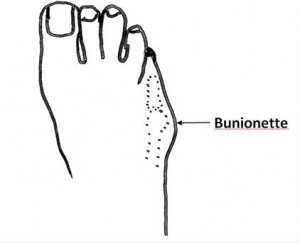
A Bunionette is a deformity at the base of the fifth or little toe (Figures 1 & 2), which tends to occur gradually over time. Patients with bunionette deformities experience localized pain on the outside aspect of the base of the fifth toe, there may also be callosity and pain where the fifth toe rubs on the forth toe.
Figure 1: Bunionette Deformity |
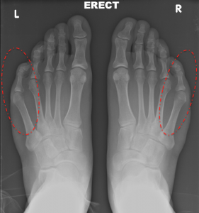
Figure 2: Pre-op x-ray |
 |
 |


Physical Examination
Physical examination will show a deformity of the fifth toe with a prominence on the outside at the base. There can be swelling in this area representing a bursa, which is a fluid-filled sac. This swelling may change in size from day to day due to inflammation, often aggravated by tight shoes. The fifth toe itself usually has a normal range of motion. The sensation and blood supply of the foot is typically normal. Sometimes, patients also have a bunion of the big toe.
Imaging Studies
Standing plain x-rays show the bony alignment that is responsible for the observed deformity. There is usually an angular deformity at the fifth metatarsophalangeal joint, which corresponds to the base of the fifth toe. In some patients, the problem is a metatarsal bone (the long bones of the mid-foot) that angles outward as well as the toe leaning inward.
Treatment
Non-Operative Treatment
Most of the cases of bunionette deformity can be managed nonoperatively. Non-operative treatment includes:
- Use of comfort shoes: Shoes with a wider toebox and comfortable soft leather upper can be helpful in managing this deformity. Occasionally, it is beneficial to have a shoemaker create extra space in this area to accommodate the deformity.
- Comfort pads: Pads can be applied over the outside aspect of the bunionette deformity to make this area more comfortable. For example, a C-shaped pad can help protect the prominent area. However, applying a pad may necessitate creating increased space within the front of the shoe.
- Toe spacer: A toe spacer can be placed between the fourth and fifth toe to help reduce the deformity and improve the patient’s symptoms.
- Activity modifications: If the deformity is severe enough, it may be necessary to limit the amount of time that the individual stands or walks.
Operative Treatment
In a small percentage of patients, operative treatment will be beneficial. Surgery should only be considered after nonoperative management has failed. The type of operative treatment is dependent upon the actual deformity. Operative options for treating bunionette deformities include:
- Removal of the prominent bone on the outer aspect of the fifth metatarsal head: In some individuals with a bunionette deformity, the outside aspect of the fifth metatarsal head is prominent and simply removing this will help the symptoms.
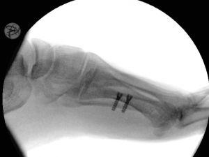
- Cutting and repositioning the fifth metatarsal bone: In most patients with significant bunionette deformities, there is an increased angle between the fourth and the fifth metatarsal with the subsequent increased deformity at the fifth toe (metatarso-phalangeal) joint. In these individuals, a formal cutting and repositioning of the bone (osteotomy) of the fifth metatarsal is necessary. Depending upon the exact nature of the deformity, the bone can be cut close to the bunionette or further up the foot to reposition the entire bone. These procedures are combined with a soft tissue tightening of the outer (lateral) joint capsule of the fifth metatarsophalangeal joint, further correcting the deformity.
Figure 3: Bunionette Lateral x-ray post-op |
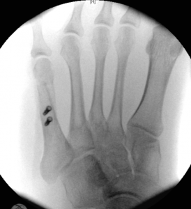
Figure 4: AP x-ray post-op |
 |
 |


Potential Operative Complications
Potential post-operative complications include general surgical complications such as:
Specific complications associated with a bunionette correction include:
- Recurrence of the Deformity
- Continued (or even increased) local pain
- Vascular Injury (including potential loss of blood supply to fifth toe)
Edited on November 24, 2018
(Previously edited by Justin Greisberg, MD and Robert Leland, MD)
mf/ 7.24.18