

Pilon Fracture
(Tibia Plafond Fracture)
Edited by Eric Malickey, MD
Summary
A Pilon fracture is a severe injury involving the ankle joint. The fracture involves the larger bone of the lower leg (tibia), and extends into the weight bearing surface of the ankle joint. It usually occurs following a significant force to the foot, such as a fall from a height or motor vehicle accident (MVA). The bone of the lower part of the ankle joint (talus) is driven into the top of the ankle joint, causing a fracture to the weight bearing portion of the ankle joint (tibial plafond) and the distal tibia (and possibly fibula). It is a painful injury associated with a fair amount of swelling. Treatment is often surgical if the fracture is displaced. Surgery attempts to restore the shattered bone back to its original position. It often takes many months for the fracture to heal and more than a year for the patient to reach their point of maximal improvement. Some ankle arthritis and joint stiffness is common even with optimally treated Pilon fractures.
Clinical Presentation
Patients with a Pilon fracture usually have suffered an excessive load or force to the ankle joint, such as a fall from a height or a motor vehicle accident. It is common for patients to have other injuries. A Pilon fracture is associated with marked pain, ankle swelling, and distorted anatomy. The injury is often extremely uncomfortable and patients cannot walk on the affected limb.
Physical Examination
Physical examination will show significant swelling involving the ankle joint and the lower leg. There may be associated deformity if the ankle fracture is significantly out of the normal position. It is important to check the nerve and the blood supply to the foot, as this can be injured. It is also important to check the other muscles, bones, and joints to see whether there are any other associated injuries. There is limited muscle and soft tissue coverage over the ankle joint, and it is not uncommon for this injury to be associated with bone fragments cutting through the skin – an open fracture. If this occurs, it is necessary to get this treated surgically in an expedited manner in order to minimize the risk of infection.
Imaging Studies
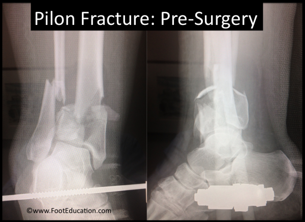
A plain x-ray will usually allow the injury to be diagnosed, (Figure 1). It will identify the fractures that involve the lower leg bone and extend into the ankle. An additional study (computerized tomography or CT scan) is usually required to elaborate the fracture pattern and to help the surgeon understand the nature of the fracture, which varies considerably. The defining feature of a Pilon fracture is a fracture that extends into the weight-bearing portion of the ankle joint.
Figure 1: Pilon Fracture before surgery

Treatment
Non-Operative Treatment
Occasionally, either because the fracture is nondisplaced (not shifted) or because surgery is contraindicated (too risky), the patient can be treated without surgery. Non-operative treatment requires a prolonged period (10 to 12 weeks) of non-weight-bearing to allow the fracture to heal. Patients are usually placed in a splint and cast during that time and use crutches.
Operative Treatment
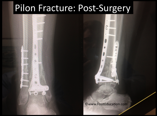
Most Pilon fractures in otherwise healthy individuals are surgically treated. This is often performed once the swelling has settled. This means that the surgery will be performed a number of days or even weeks after the original injury. During this time, the foot will be elevated and sometimes it will be placed in an external frame (external fixator) to help stabilize the soft tissues that are often significantly damaged after this type of injury. The surgery itself can be quite complex. It essentially involves repositioning the fractured bone fragments back into the right position. It is particularly important that the bone that makes up the top part of the ankle joint (the tibial plafond) be restored to as close to a normal (anatomic) position as possible. This surgical procedure is performed through several skin incisions. Once the fractures are provisionally fixed with wires, they are definitively stabilized with plates and screws (Figure 2). Sometimes the smaller bone located on the side of the ankle joint (the fibula), is also fractured and requires fixation.
Following the surgery, the patient is kept non-weight bearing until the bone heals, which often takes 10 to 12 weeks or more. During this time the patient uses crutches, a knee walker, and a wheelchair to mobilize.
Figure 2: Pilon Fracture Post-Surgery

Potential Complications
This is a potentially very serious high-energy injury with a reasonably high complication rate. The main surgical complications are:
- Infection
- Wound healing issues
- Painful prominent hardware. This may also be a complication over time. In some instances, the hardware will need to be removed after the fracture has healed.
- Ankle Arthritis. Many patients who suffer a Pilon fracture will develop some post-traumatic ankle arthritis.
Edited September 23, 2017
Previously edited by Hossein Pakzad, MD
mf/ 5.29.18