

Metatarsalgia
Summary
Watch Video: Metatarsalgia

Metatarsalgia is characterized by pain in the forefoot. The term literally means “pain on the metatarsal” (there are 5 metatarsal bones in the forefoot). Metatarsalgia is not a true diagnosis, but rather it is a symptom. Patients with metatarsalgia present with pain in their forefoot, usually in the ball of the foot (Figure 1). The pain is often described as aching, and it is typically aggravated by standing and walking. In general, metatarsalgia is caused by repetitive overloading of the forefoot, leading to chronic localized tissue injury. Often the most symptomatic area is at the base of the 2nd or 3rd toe. Factors that may predispose to the development of metatarsalgia include: a bunion deformity, arthritis of the great toe, ligament instability of the midfoot, an excessively tight calf muscle, a congenital foot deformity, and claw toe deformities.
Non-operative treatment of metatarsalgia is often successful. Treatment principles include:
- Making the correct diagnosis and addressing the underlying cause of the symptom.
- Diminishing the repetitive loading through the forefoot.
- Dispersing the loading on the forefoot over a wider area.
Click here for a detailed summary handout on Metatarsalgia
Figure 1: Typical Pain Location
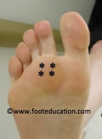

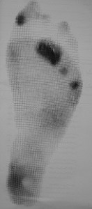
Figure 2: Localized forefoot load

Clinical Presentation
The pain that is experienced in metatarsalgia typically occurs in the ball of the foot (at the base of the second or third toes under the second or third metatarsal heads). It often feels like walking “on stones or pebbles” or on “a rolled up sock” in the front of the foot. The pain is often a deep ache and it is worse with walking on hard surfaces barefooted. There may also be a burning feeling extending into the tips of the second and third toes.
As the condition progresses, it may be associated with increased curling of the toes (clawtoes). This causes the padding of the forefoot to be pulled away and results in greater pressure beneath the bones of the foot. The result is a loss of protection from the metatarsal heads, less shock absorption with walking, callus formation, and more pain in the foot
Metatarsalgia can be confused with other “ball of foot conditions.” Many patients will have “clawtoes,” which is a curling of the little toes. Both conditions often occur and can be described by providers as the cause of the pain.
If an ink imprint of the weight bearing sole of the foot is obtained, such as with a Harris mat (a device that assesses how force is distributed throughout the foot), there is often an intense uptake in the area corresponding to the involved metatarsal head(s) (Figure 2).
Continued, localized, repetitive walking will predispose to chronic injury to the structures of the forefoot. The structures that are commonly injured include the MTP joint capsule, the plantar plate, and the metatarsal bone (head and/or neck). The irritation of the MTP joint is due to repetitive overload and can lead to swelling of the joint itself (Capsulitis or MTPJ Synovitis). The second and third toes may separate in metatarsalgia and patients will either have pain at the bottom and/or top of the ball of the foot (base of the toes).
Note: Many patients and physicians misdiagnose a Morton’s neuroma for variants of metatarsalgia. In general, metatarsalgia is more common in the second and third toes whereas Morton’s neuroma involves the third and forth toe area. Also metatarsalgia generally hurts more when barefooted, whereas Morton’s neuroma hurts in tight fitting shoes.
Imaging Studies
X-rays in patients with metatarsalgia often demonstrate a long second or third metatarsal, relative to the first and the fourth metatarsal. In rare instances, the MTP joint may actually be partially or completely out of joint (subluxed or dislocated). Deformities of the 1st metatarsal, such as those present with a Bunion or with midfoot instability, may also be observed on x-ray.
Treatment
Non-Operative Treatment
Patients respond well to non-operative treatment. the goal of non-surgical treatment is to take weight off the painful area of the foot. This can be done with a combination of soft padded comfortable shoes, metatarsal pads, soft accommodative orthotics, activity modifications (avoid putting forced pressure to the ball of foot), calf stretching, foot muscle strengthening (try and gently curl toes), and NSAIDS (Non-steroidal anti-inflammatory drugs).
- Comfort shoes: Unfortunately, high heels and pointed toe shoes will worsen the symptoms, by concentrating weight bearing to the ball of the foot. Stiff sole/rocker bottom shoes with soft liners can help lessen pressure to the ball of the foot. Orthopaedic shoes of the past carry a negative connotation of an ugly, unstylish, men’s Oxford, which are rejected by many patients. Currently, many women’s and men’s dress and athletic shoe manufacturers carry lines with more style and the same features. As a rule, “if the shoe fits” and feels good, it is probably fine to use.
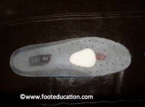
- Metatarsal pads: Metatarsal pads can be very helpful in treating metatarsalgia (Figure 3). When metatarsal pads are fitted appropriately, they will shift weight away from the metatarsal heads that are involved. As noted in Figure 3, to be effective a metatarsal pad needs to be positioned before the main area of loading. If the pad makes the foot hurt more, it is likely too far forward in the shoe. See also: How do I fit a Metatarsal Pad?
Figure 3: Metatarsal Pad

- Soft Orthotics: Soft accommodative orthotics help to cushion the ball of the foot. Look for a reasonably priced over the counter soft padded shoe liner or a gel insert.
- Slight heel rise: For some patients, a slight rise in the heel of the shoe may actually lessen pain in the ball of the foot. This is because many folks will have a tight calf muscle (equinus) which tends to result in more pressure beneath the ball of the foot. The heel lift helps shift weight back to the heel pad sooner (and offloads the ball of the foot).
- Hammertoe crest pad: For patients with an associated clawtoe deformity, it may be helpful to use a hammertoe crest pad, or toe taping, to help bring the toe back into an improved position. This may serve to help pull the toe down and in turn restore the plantar fat pad under the metatarsal head.
- Activity modification: A calf muscle contracture (equinus contracture) will increase stress on the forefoot. Stretching the calf will help eliminate this cause of pain, but patient’s must stretch the calf on an incline board wearing shoes and avoid concentrating the body weight through the ball of the foot.
- Non-steroidal anti-inflammatory drugs: NSAIDs can be very helpful if symptoms are moderate or severe, as they can modify the perception of symptoms, giving other non-operative treatments time to allow the overloaded area to heal. NSAIDs are most effective being in the body over a 10-14 day period therefore consult your physician before starting.
- Corticosteroid Injection: Injecting corticosteroids into the involved joint (MTPJ) can give temporary relief (1-3 months) in certain cases but beware. Cortisone may weaken the toe joint restrains (Plantar plate disruption). If this occurs, the toe can become more deformed (worsening clawtoe) and the metatarsalgia will become much worse.
Operative Treatment
In a small percentage of patients surgery is needed for metatarsalgia. There are a variety of procedures that have been proposed, either in isolation or in combination to address Metatarsalgia. These include shortening the bones of the foot (corrective osteotomy), releasing the calf muscle (correcting the equinus contracture) and/or correcting the clawtoe deformity. It is essential that the primary cause of the metatarsalgia be addressed. The technique chosen will depend on the extent of the deformity, the stiffness of the toe, and the preference of the surgeon.
If the joint at the base of the involved toe (MTP joint) is swollen and inflamed it may be helpful to remove the inflamed synovial lining (synovectomy). This is often done in conjunction with other procedures. If the second and/or third metatarsal heads are “long,” they may be shortened to equal the weight through the ball of the foot.
If a Bunion deformity is present, this may need to be corrected in order to address the abnormal weight bearing of the forefoot. If a tight calf muscle is present and does not resolve with stretching, lengthening of the gastrocnemius muscle (Strayer procedure) may be beneficial.
General Potential Complications
The usual list of general post-surgical complications may occur with various procedures that are used to address metatarsalgia. This includes the potential for:
- Wound healing problems
- Infection
- Nerve injury to the local nerves that provide sensation to the tips of the toes
- Deep Vein Thrombosis (DVT) – uncommon
- Pulmonary Embolism (PE) – very uncommon
Specific Complications
Surgery on the toes and forefoot is not as predictable or as easy as patients think. Specific complications depend on the procedure(s) that are performed but can include:
- Continued symptoms: It is often difficult to eliminate all of the pain because metatarsalgia is typically a long-term problem. There is a certain amount of tissue damage that has already occurred, which can’t be reversed.
- Stiffness of the toe: Toes that have been operated on lose some flexibility. Usually this is not a major problem, but in some instances it can lead to discomfort.
- Recurrent deformity of the toe: Toe surgery is performed in an attempt to correct or improve the deformity and associated symptoms. However, balancing and positioning the toe can be tricky, and a recurrent cock-up deformity of the toe can occur. This is frustrating to the patient and physician.
- Numbness in part or all of the toe: Although unlikely, this occurs because the small nerves lie close to the sides of the toe. Numbness can take a year to reverse.
- Transfer metatarsalgia: Surgery that corrects metatarsalgia changes the loading characteristics of the front of the foot. In some unusual cases this may lead to increased pressure (pain) else where in the foot.
- Loss of blood supply to the tip of the toe: In extremely rare cases, the blood supply to the tip of the toe is completely stopped. There are two small arteries (one on either side of the toe) that supply blood to the tip of the toe. If the blood supply to the tip of the toe is lost the tissue will die and it may be necessary to remove part or all of the toe.
Click here for a detailed summary handout on Metatarsalgia
Previously Edited by: Eric Malicky, MD, Vinod Panchbhavi, MD
Edited on November 1, 2018
mf/ 06.11.18