

Calcaneal Fracture ORIF
Edited by David Garras, MD
Indications
The term “ORIF” stands for “open reduction, internal fixation.” Quite simply, the surgeon opens the fracture site, puts the fractured bone back together then, uses plates and/or screws to stabilize the broken bone pieces. The main indication for this type of surgery on the heel bone (calcaneus fracture) is a displaced fracture. Displaced means the bone fragments are “out of position” and must be put back together. A calcaneus fracture (heel bone) commonly includes damage to the subtalar joint. This is the joint that allows side-to-side motion of the foot. A damaged joint can lead to arthritis. By putting these broken pieces back together, orthopaedic surgeons hope to maximize the functional outcome and reduce the chance that arthritis will develop. However, not all patients with calcaneal fractures are candidates for surgery. Certain fractures are not displaced enough to need surgery. And for other patients, the risk of a serious complication (such as a deep infection) may outweigh the potential benefits of surgery. It is often the judgment of the surgeon (after discussion with the patient) to determine the best treatment method.
Procedure
The goal of calcaneal fracture ORIF is to place the bones back to their original position prior to the injury. By restoring the normal alignment, the surgeon hopes to provide the patient with the best possible outcome. The final outcome often depends on the severity of the initial injury. Simple fracture patterns with minimal displacement and large bone pieces are easier to treat than highly complex fractures involving many small pieces. During surgery, these small pieces all need to be carefully placed in their original position. This challenging endeavor is equivalent to putting a broken eggshell back together.
During surgery, the patient is usually positioned on his or her side. The incision is made on the outside (lateral) aspect of the foot. Your surgeon will determine the type and shape of this incision. Some fractures can be approached through small incisions and others require longer incisions. Once the skin incision is made, your surgeon will carefully move important tendons, nerves, and ligaments out of the way in an effort to visualize the underlying calcaneus bone. Sometimes these tendons, nerves, and ligaments are damaged along with the calcaneal bone and need to be repaired too. Once the bone is exposed, your surgeon will make an effort to inspect the subtalar joint. The subtalar joint, which is often injured in calcaneus fractures, is the joint below your ankle that controls the “side-to-side” (inversion and eversion) motion of your foot.
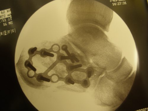
Once the surgical exposure is complete, the process of systematically reassembling the bone pieces is started. There are usually two primary fragments of the fractured calcaneus. These fragments are realigned first and temporarily fixed with wires to hold them in place. Proper alignment is confirmed by the use of a live x-ray machine (called a C-arm). Next, the subtalar joint surface is reconstructed by systematically repositioning all other fracture fragments. Once the calcaneal alignment is restored, the temporary wires are sequentially removed and replaced with permanent hardware, such as plates and/or screws (Figure 1A and 1B).
The final step involves closure of the surgical wound. In some ways, this is the most important part of the surgical procedure because any breakdown of the wound will increase the risk of a significant post-operative infection. Once the wound closure is complete, a soft cast (called a splint) is applied to the leg.
Figure 1A: Fractured Calcaneus
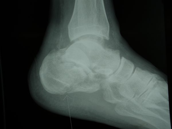

Figure 1B: Fixed Calcaneal Fracture
Recovery
Week 0-2 Post Surgery
The foot is immobilized in the soft cast (splint), iced and elevated. Your surgeon will ask that you not place any weight on your foot (non weight-bearing). This will require the use of a wheelchair, walker, knee scooter and/or crutches. You will likely require prescription-strength pain pills. Over-the-counter laxatives and stool softeners may be required to prevent or treat constipation. Your dressing should remain clean and dry. Do not change your dressing unless instructed by your surgeon.
Week 2-6 Post Surgery
At your first post-operative visit, your doctor will remove your splint and examine your incision. Sutures may need to be removed. You will be placed either into a cast (which can’t be removed) or a removable boot. For most calcaneus fractures, your surgeon will ask that you remain non weight-bearing until the fracture demonstrates adequate healing – as seen on subsequent x-rays. If you are placed in removable boot, your surgeon may ask that you start a gentle range-of-motion program.
Week 6-12 Post Surgery
During this time period, you are working to improve the range of motion of your foot and ankle. This may involve visits to a physical therapist combined with a home exercise program. Your surgeon may also allow you to gradually increase your weight-bearing forces. At the end of this time period, if the fracture shows evidence of solid healing, your surgeon may allow you to transition out of the boot and possibly into an ankle brace. The ankle brace will require the use of a wide, stable and comfortable shoe.
Week 12-24 Post Surgery
Once the bone has completely healed, you will begin more advanced physical therapy activities (such as walking without a limp and improving your balance and strength). You will see a gradual reduction of swelling, although expect your foot/ankle to remain swollen for many months after surgery. Full recovery from these challenging injuries often takes 12 months or more.
Potential General Complications
- Asymmetric Gait (leading to pain elsewhere)
- Deep Vein Thrombosis
- Failure to Resolve ALL Symptoms
- Pulmonary Embolism (PE)
Potential Specific Complications
- Wound Healing Problems: Although seen with any surgical procedure, wound healing complications are particularly concerning following calcaneal fracture surgery. The area around the outside of the heel has relatively thin skin and limited soft-tissue coverage. This can make wound healing problems more likely following calcaneal fracture surgery, and potentially more severe if they do develop. Wound healing problems are increased significantly for smokers and diabetics.
- Infection: Infections can create a major problem if they occur following a calcaneal fracture. As a result of the limited soft-tissue covering the outside of the heel, a superficial wound infection can quickly spread down to the underlying bone. If an infection develops, your surgeon may recommend the use of oral or intravenous antibiotics. A repeat trip to the operating room may be required.
- Sural Nerve Injury: Injury to the nerve on the outside of the heel (sural nerve) can occur during calcaneal fracture surgery. Nerve injury can occur due to retraction, direct injury, or from scarring during the recovery process. If the sural nerve is injured or cut, the patient may experience numbness or pain along the path of the nerve.
- Subtalar Arthritis: Painful subtalar arthritis and stiffness of the hindfoot is common following calcaneal fractures. This occurs as a result of the damage to the cartilage at the time of the initial injury.
- Painful Hardware: Pain may be associated with the screws and plates used to align and secure the broken bone fragments. This occurs in about 10-20% of patients who have had surgical stabilization of a calcaneus fracture. Your doctor will help you determine if hardware removal is required.
Previously Edited by Steven Neufeld, MD and Matthew Buchanan, MD
Edited September 29, 2019
mf/2.6.18